


Evaluation of the prosthetic space in completely edentulous Moroccan patients: a cross-sectional study
##plugins.themes.academic_pro.article.sidebar##
##plugins.themes.academic_pro.article.main##
Abstract
Introduction: In completely edentulous patients, the prosthetic space corresponds to the area available for denture integration. Its evaluation relies on inter-ridge height, ridge dimensions, and the degree of alveolar resorption, which condition the functional and esthetic outcome of prosthetic rehabilitation.
Objective: To assess prosthetic space parameters in completely edentulous Moroccan patients and identify associated factors.
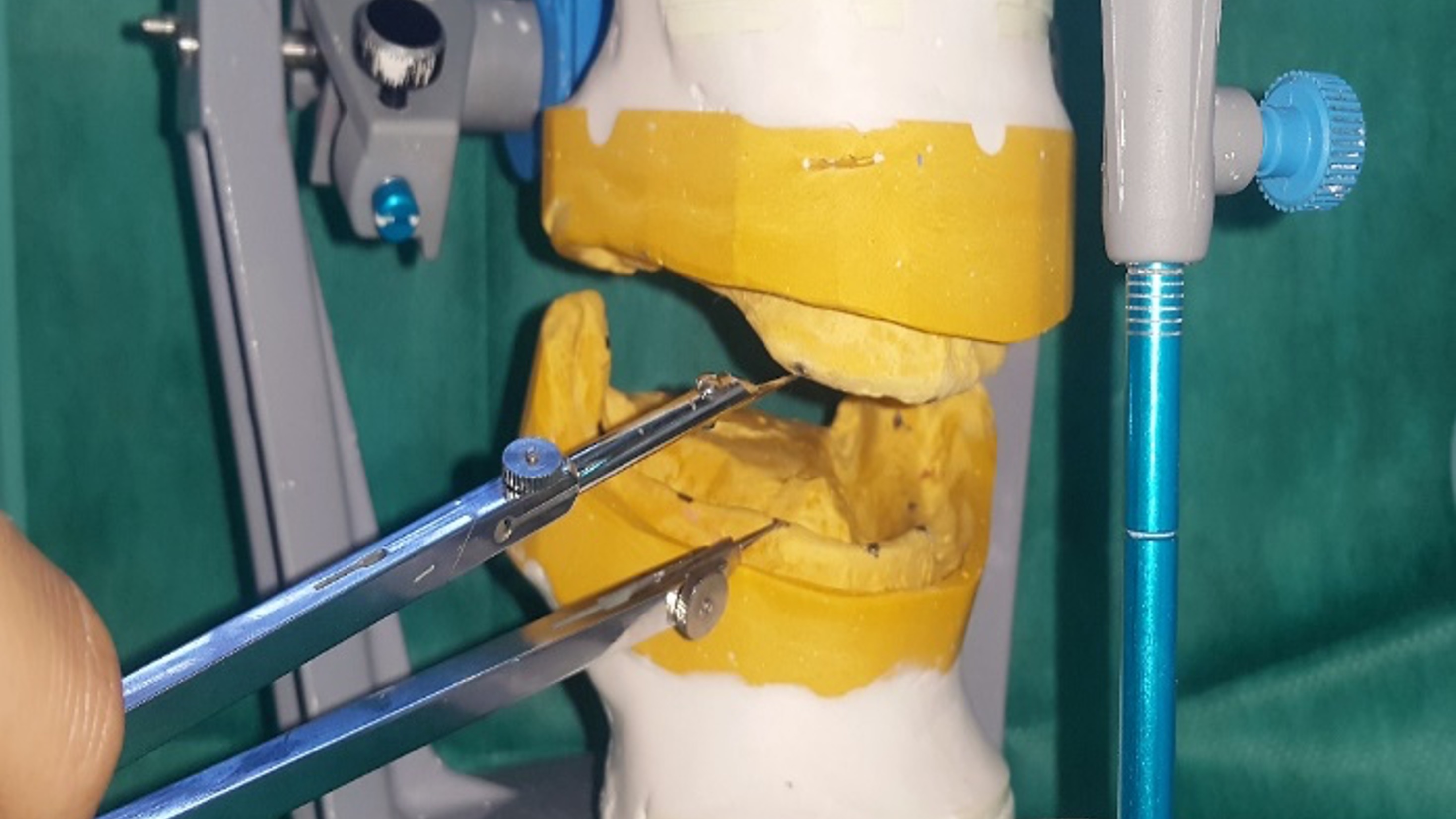
Methods: A cross-sectional study was conducted on 50 completely edentulous patients consulting for bimaxillary rehabilitation at Ibn Sina Hospital, Rabat. After recording the maxillomandibular relationship, secondary casts were mounted on an articulator to measure inter-ridge height, ridge dimensions, and arch perimeters. Maxillary and mandibular resorption were classified using Cawood and Howell's classification. Data were analyzed with Jamovi 2.6.13.
Results: Inter-ridge height was greater at the median level [2 cm (1.7–2.2 cm) right; 1.95 cm (1.7–2.3 cm) left] and lower at the tuberosity-trigone level [0.95 cm (0.7–1.48 cm) right; 1.05 cm (0.8–1.48 cm) left] (p < 0.001). Maxillary resorption was predominantly classified as type III, whereas mandibular resorption mainly corresponded to classes IV and V, both anteriorly and posteriorly (p < 0.001). Age was significantly associated with anterior mandibular resorption (p < 0.05), and the duration of edentulism correlated with resorption in both arches (p < 0.05).
Conclusion: The joint evaluation of inter-ridge height, ridge perimeters and dimensions, as well as resorption profiles allows for an objective and comprehensive analysis of prosthetic space in edentulous patients.
Keywords:
prosthetic space, inter-ridge height, alveolar resorption, gender, age, duration of edentulism##plugins.themes.academic_pro.article.details##

This work is licensed under a Creative Commons Attribution-NonCommercial-NoDerivatives 4.0 International License.
References
- Ahuja S, Cagna DR. Defining available restorative space for implant overdentures. J Prosthet Dent.2010;104(2):133-6.
- Heath MR. Facteurs cliniques qui déterminent l’espace prothétique. Congrès ADF 1972.Inf Dent. 1973;1:70-1.
- Klein P. Quelques définitions pour mieux comprendre la piézographie.Inf Dent.1989; 71(29):2519-23.
- Nabid A. Traité odontologique de piézologie. Alger: ENAG Editions; 2014. 592 p.
- Kordatzis K, Wright PS, Meijer HJA. Posterior mandibular residual ridge resorption in patients with conventional dentures and implant overdentures. Int J Oral Maxillofac Implants. 2003;18(3):447-52.
- Hirai T, Ishijima T, Hashikawa Y, Yajima T. Osteoporosis and reduction of residual ridge in edentulous patients. J Prosthet Dent. 1993;69(1):49-56.
- Atwood DA. Some clinical factors related to rate of resorption of residual ridges. J Prosthet Dent. 2001;86(2):119-25.
- Atwood DA. Bone loss of edentulous alveolar ridges. J Periodontol. 1979;50(4 Spec No):11-21.
- Soikkonen K, Ainamo A, Xie Q. Height of the residual ridge and radiographic appearance of bony structure in the jaws of clinically edentulous elderly people. J Oral Rehabil. 1996;23(7):470-5.
- de Baat C, Kalk W, van ’t Hof M. Factors connected with alveolar bone resorption among institutionalized elderly people. Community Dent Oral Epidemiol. 1993;21(5):317-20.
- Nishimura I, Hosokawa R, Atwood DA. The knife-edge tendency in mandibular residual ridges in women. J Prosthet Dent. 1992;67(6):82-6.
- Xie Q, Ainamo A, Tilvis R. Association of residual ridge resorption with systemic factors in home-living elderly subjects. Acta Odontol Scand. 1997;55(5):299-305.
- Frankenthal S, Nakhoul F, Machtei EE, Green J, Ardekian L, Laufer D, et al. The effect of secondary hyperparathyroidism and hemodialysis therapy on alveolar bone and periodontium. J Clin Periodontol. 2002;29(6):479-83.
- Imirzalioglu P, Yuzugullu B, Gulsahi A. Correlation between residual ridge resorption and radiomorphometric indices. Gerodontology. 2012;29(2):e536-42.
- Bairam LR, Miller WA. Mandible bone resorption as determined from panoramic radiographs in edentulous male individuals ages 25-80 years. Gerodontology. 1994;11(2):80-5.
- AlSheikh HA, AlZain S, Warsy A, AlMukaynizi F, AlThomali A. Mandibular residual ridge height in relation to age, gender and duration of edentulism in a Saudi population: A clinical and radiographic study. Saudi Dent J. 2019;31(2):258-264.
- Parr NM, Passalacqua NV, Skorpinski K. Investigations into Age‐related Changes in the Human Mandible. J Forensic Sci. 2017;62(6):1586-1591.
- Bressan E, Ferrarese N, Pramstraller M, Lops D, Farina R, Tomasi C. Ridge Dimensions of the Edentulous Mandible in Posterior Sextants: An Observational Study on Cone Beam Computed Tomography Radiographs. Implant Dent. 2017;26(1):66-72.
- Cawood JI, Howell RA. A classification of the edentulous jaws. Int J Oral Maxillofac Surg. 1988;17(4):232-6.
- Alidema SH, Bundevska J, Bimbashi V, Maja S, Dimoski G, Halili R. Cone Beam Computed Tomography (CBCT) Evaluation of Bone Height and Width in Edentulous Mandible. Int J Biomed. 2022;12(3):417-422.
- Pietrokovski J, Starinsky R, Arensburg B, Kaffe I. Morphologic Characteristics of Bony Edentulous Jaws. J Prosthodont. 2007;16(2):141-7.
- Pramstraller M, Schincaglia GP, Vecchiatini R, Farina R, Trombelli L. Alveolar ridge dimensions in mandibular posterior regions: a retrospective comparative study of dentate and edentulous sites using computerized tomography data. Surg Radiol Anat. 2018;40(12):1419-1428.
- Huumonen S, Haikola B, Oikarinen K, Söderholm AL, Remes-Lyly T, Sipilä K. Residual ridge resorption, lower denture stability and subjective complaints among edentulous individuals. J Oral Rehabil. 2012;39(5):384-90.
- Tayachi Y, Tayari O, Bendhief R, Jaouadi J. Bone Resorption Evaluation According to Measurements’ Panoramic Radiographs: A Tunisian Cross-Sectional Study. Saudi J Oral Dent Res. 2021;6(11):511-518.
- Koç A, Erdem R. Evaluating Edentulous Mandible Alveolar Crest Dimensions in Posterior Region By Cone Beam Computed Tomography. East J Med. 2020;25(3):345-349.